

As a leading cataract surgeon in Houston, I have the opportunity to see many patients with many different types of cataracts. At the time that I diagnose cataracts in the clinic, often patients will ask, “What is a cataract and what does it look like? Is it a film over my eyes?”.
Well, not really. The front clear window on the eye is the cornea. Under the cornea is the colored part of the eye called the iris. Behind the iris is the lens which is normally clear. By the age of 40, most people have at least the beginning of a clouding and yellowing of the lens which we call a cataract.
You’ve heard the phrase a picture is worth a thousand words…….
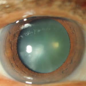
Cataract before cataract surgery
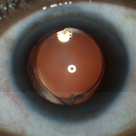
Intraocular lens (IOL) after cataract surgery
We normally remove cataracts with phacoemulsification which is very high frequency ultrasound that breaks up the cataract into pieces and simultaneously vacuums it out.
Here is a video that shows a patient having their scarred cornea removed (for a corneal transplant) followed by the cataract being removed. It allows you to get a real sense of the size and color of a cataract: Rare surgical view of whole intact cataract