

There are many types of eye tests to detect problems. Ophthalmologists have to find the problems before we can know what treatments will work best.
Cornea Tests
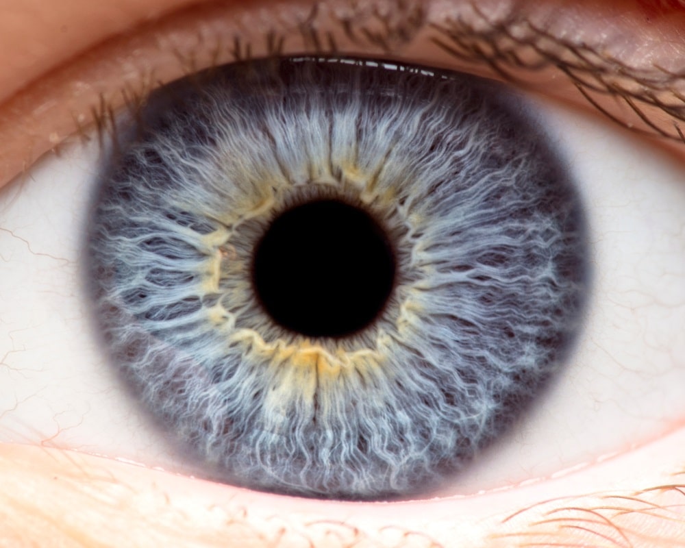
Photos: used to document the appearance, size, and location of eye structures in the front of the eye.
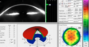
Corneal Topography: used to map the clarity, shape, curves, and thickness of the cornea.
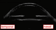
Corneal OCT: used for in depth details of the structure of the cornea.
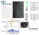
Endothelial Cell Count: used to measure the number, size, and shape of the cells that keep the cornea clear and compact.
Corneal Pachymetry: used to measure the thickness of the cornea.
Lens Tests
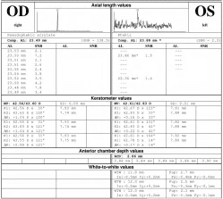
Intraocular Lens (IOL) Measurements: used to determine the correct power of the lens inserted during cataract surgery.
Anterior segment UMB or OCT: used to examine the structures in the front of the eye when the cornea is not clear.
Retina Tests
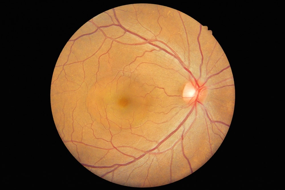
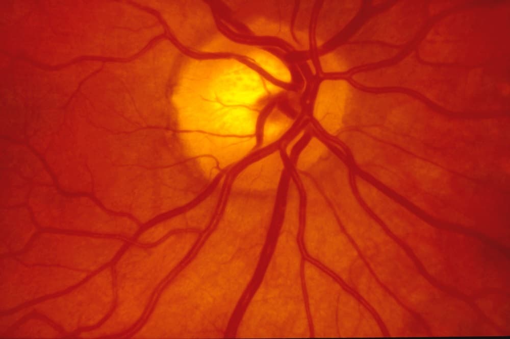
Photos: used to document the appearance, size, and location of structures in the back of the eye.
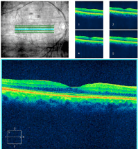
Retinal OCT: used for in depth details of the structure of the retina.
Flourescein Angiography: used to map the flow and leakage of blood in the vessels of the retina.
Optic Nerve Tests
Photos: used to document of appearance, size, and location of the optic nerve.
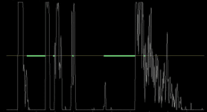
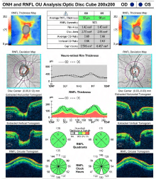
Optic Nerve OCT: used for in-depth details of the structure of the optic nerve.
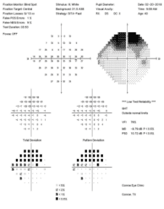
Visual Field: used to map any problems with central or peripheral vision.
Dr. Vital’s office at Houston Eye Associates has the latest technology in ocular testing to help you see better.